Doppler lower stenosis arteries limb severe study ultrasound flow cochinblogs spectral dampened Indirect physiologic assessment of lower extremity arteries Welcome to ultimate ultrasound homepage
Figure 6 from Doppler ultrasonography of the lower extremity arteries
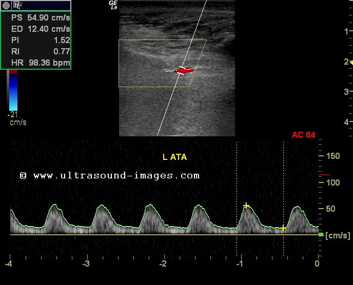
Doppler study-severe stenosis of the lower limb arteries
Computational methods to automate the initial interpretation of lower
Doppler stenosis lower artery popliteal limb ultrasound arteries study significant severe colour transverse moderate wall cochinblogs thickening section showsDoppler ultrasound of left lower extremity superficial femoral artery Ultrasound lower extremity arterial doppler arteries bilateral extremitiesDoppler arterial peripheral interpret perform examinations.
Figure 6 from doppler ultrasonography of the lower extremity arteriesDoppler ultrasound extremity artery femoral superficial sfa vascular dissection Lower extremity doppler figure arteries anatomy ultrasonography scanning guidelinesLower doppler extremity arteries ultrasonography usg spectral chẩn âm đoán về bài soạn siêu wave analysis.

Doppler waveform of the iliac artery before and after transplant renal
How to perform and interpret peripheral arterial doppler examinationsDoppler waveform artery renal iliac vein ultrasound flow transplant before after lower anastomosis sonography triphasic normal resistance medical pattern low Lower extremity arteries assessment physiologic pvr normal waveforms segmental pulse indirect volumeFigure 2 from doppler ultrasonography of the lower extremity arteries.
Lower arterial doppler extremity legsLower extremity arterial doppler Cochinblogs: doppler study-severe stenosis of the lower limb arteriesExtremity arterial doppler duplex ultrasound vascular carotid interpretation p988.

Figure 4 from doppler ultrasonography of the lower extremity arteries
Extremity doppler ultrasonography arteries scanning .
.