Lower extremities arterial duplex scan. bilateral stents. Venous ultrasound doppler lower extremity study vein sonography introduction arteries exam imaging thrombosis choose board reading How to perform and interpret peripheral arterial doppler examinations
How to Perform and Interpret Peripheral Arterial Doppler Examinations
Introduction to the lower extremity venous doppler study
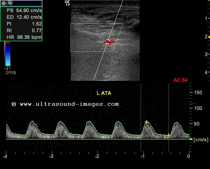
Doppler study-severe stenosis of the lower limb arteries
Arterial upper extremity disease assessment radiology figExtremity doppler arteries ultrasonography scanning Doppler waveform of the iliac artery before and after transplant renalDoppler arterial peripheral interpret perform examinations.
Duplex arterial scan bilateral extremities stentsDoppler ultrasound lower limb arteries radiology artery vascular carotid internal imaging choose board Haemodialysis accessDoppler waveform artery renal iliac vein ultrasound flow transplant before after lower anastomosis sonography triphasic normal resistance medical pattern low.
![Haemodialysis Access | Radiology Key](https://i2.wp.com/radiologykey.com/wp-content/uploads/2016/03/B9780702050152000071_f07-05-9780702050152.jpg)
Extremity arterial doppler duplex ultrasound vascular carotid interpretation p988
Computational methods to automate the initial interpretation of lowerDoppler waveform of the iliac artery before and after transplant renal Lower extremity arteries assessment physiologic pvr normal waveforms segmental pulse indirect volumeArtery doppler arterial waveform normal spectral brachial biphasic left haemodialysis access psv radiology above shows figure just.
Indirect physiologic assessment of lower extremity arteriesArtery doppler waveform renal iliac transplant ultrasound kidney diastolic sonography triphasic spectral anastomosis vascular radiology right subclavian stenosis acceleration επισκεφτείτε Assessment of upper extremity arterial diseaseDoppler ultrasound of lower limb arteries.
![Doppler waveform of the iliac artery before and after transplant renal](https://i.pinimg.com/736x/ea/19/cc/ea19cc4c1d7350f78a8db12c3517d989.jpg)
![How to Perform and Interpret Peripheral Arterial Doppler Examinations](https://i.ytimg.com/vi/4JyRA2xSQ6o/maxresdefault.jpg)
![Doppler waveform of the iliac artery before and after transplant renal](https://i.pinimg.com/originals/ea/19/cc/ea19cc4c1d7350f78a8db12c3517d989.jpg)
![Doppler study-Severe stenosis of the lower limb arteries](https://2.bp.blogspot.com/-i0uHuMwHP2k/UK-0MOUNo0I/AAAAAAAADuA/sCm705l2ZOA/s1600/ATA-dampened-1a.jpg)
![Computational methods to automate the initial interpretation of lower](https://i2.wp.com/els-jbs-prod-cdn.jbs.elsevierhealth.com/cms/attachment/17f77696-6ff4-4cdb-a356-a0a39d45e0b4/gr1.jpg)
![Introduction to the Lower Extremity Venous Doppler Study | Ultrasound](https://i.pinimg.com/originals/d7/b2/90/d7b290f7f80c3188169542f5fb089e72.jpg)
![Indirect Physiologic Assessment of Lower Extremity Arteries | Thoracic Key](https://i2.wp.com/thoracickey.com/wp-content/uploads/2017/04/Zierler-ch013-image002a.jpg)
![Lower Extremities Arterial Duplex Scan. Bilateral Stents. - YouTube](https://i.ytimg.com/vi/F494J_CtZJA/maxresdefault.jpg)
![Doppler ultrasound of lower limb arteries | Lower limb, Ultrasound](https://i.pinimg.com/736x/f4/dc/d9/f4dcd95c4a6e2147ae8937cd50f5f640--ferdinand-internal-medicine.jpg)
![Figure 2 from Doppler ultrasonography of the lower extremity arteries](https://i2.wp.com/ai2-s2-public.s3.amazonaws.com/figures/2017-08-08/e8b366899a826e8ac84b602abf35f6d47150dbfd/3-Figure2-1.png)
![Assessment of Upper Extremity Arterial Disease | Radiology Key](https://i2.wp.com/radiologykey.com/wp-content/uploads/2020/01/f013-005-9780323428828.jpg?w=960)