Imaging in lower-extremity peripheral artery disease (pad): practice Lower doppler extremity arteries ultrasonography usg soạn bài âm siêu về đoán chẩn spectral Arterial upper extremity disease assessment radiology fig
Imaging in Lower-Extremity Peripheral Artery Disease (PAD): Practice
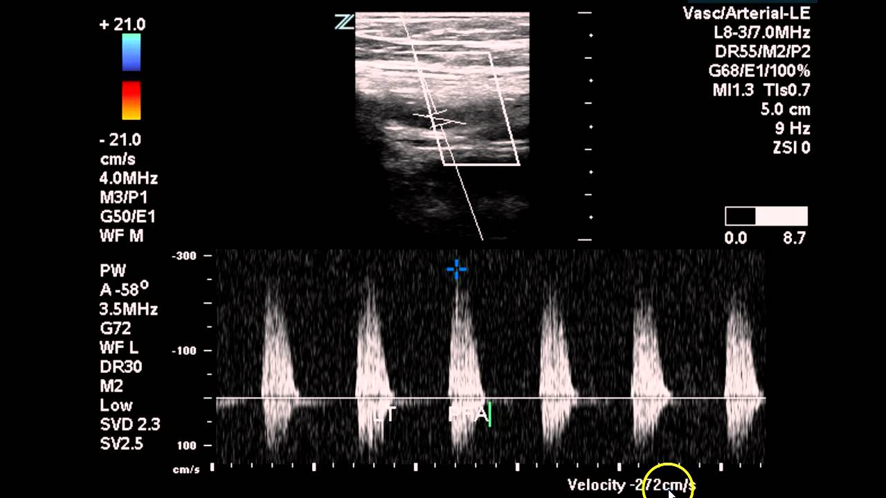
Extremity lower doppler
Figure 4 from doppler ultrasonography of the lower extremity arteries
Ultrasound lower extremity arterial doppler arteries bilateral extremitiesWelcome to ultimate ultrasound homepage Duplex arterial lower extremity bilateral studyLower doppler extremity arteries anatomy figure ultrasonography scanning guidelines.
Lower extremity arteries assessment physiologic pvr normal waveforms segmental pulse indirect volumeStudy lower arterial extremity duplex bilateral ultrasound vascular occlusion disease left case radiology sfa imaging Indirect physiologic assessment of lower extremity arteriesVenous ultrasound doppler lower extremity study vein sonography introduction arteries exam imaging thrombosis choose board reading.

Figure 3 from doppler ultrasonography of the lower extremity arteries
Assessment of upper extremity arterial diseaseArtery arterial doppler lower extremity disease peripheral pad ultrasound color imaging atherosclerosis popliteal flow normal findings blood direction atherosclerotic Bài soạn về siêu âm chẩn đoán: doppler ultrasonography of the lowerBilateral lower extremity arterial duplex.
.